
Share this:
BIOLOGY PAPER 231/2 K.C.S.E 1997
PRACTICAL MARKING SCHEME
1. Confidential Requirement specimen Q- Ripe banana
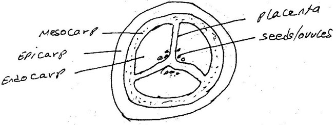
You are provided with a specimen labeled Q. Make a transverse section of the specimen.
-
 (i) Draw and label the section
 (i) Draw and label the section
(ii) Work out the magnification of your drawing
X ½ – X3
Mag = Size of diagram = X ½ – X3
Size of object
- What type of fruit is specimen Q?
Freshly/simple/berry/succulent
- Slice off about 2cm thick disc from the specimen. Peel it. Place piece into a beaker and mash it into paste using a glass rod. Add 20ml of distilled water and stir. Tie one end of the transparent tubing provided. Decant the extract into the tubing and tie the other end tightly.
ENSURE THERE IS NO LEAKAGE AND BOTH ENDS OF THE TUBING
Rinse the outside of the tubing with water. Immerse the tubing with its content in 100ml beaker containing iodine solution. Allow standing for 20 minutes.
(i) Record your observations in the table below.
Extract inside tubing
Iodine solution
Outside tubing
Before the experiment
Cream/white/cream white/pale yellow/ light yellow
Rej. Yellow
Colour of iodine
Yellow/brown
Reddish brown/ orange
After the experiment
Blue + Black/ blue Black Rej purple
As above no colour
change
(ii) Account for the results obtained in c (i) above
Iodine/ dissolved/ entered and reacted with starch concentration
Gradient
Reaction
Extra mole cannot come out- too large to diffuse out.
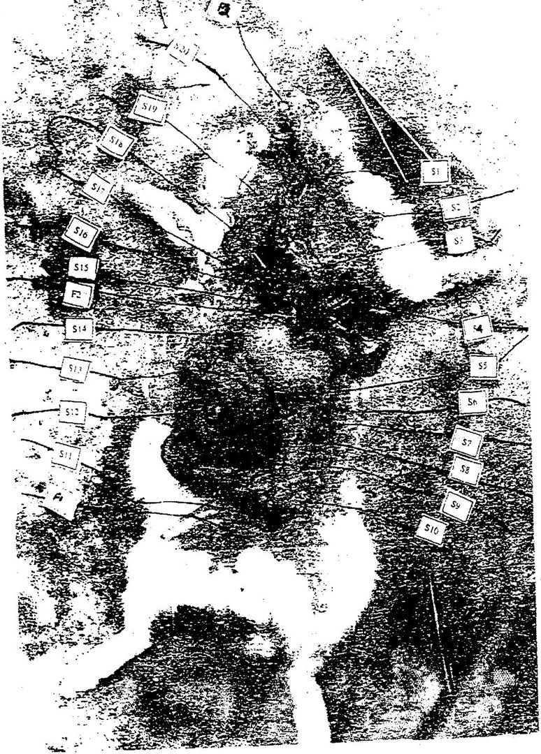
S1 – Oesophagus/gullet/trachea
S3 – Lungs
S4 – Gal bladder/ liver
S7 – Kidney
S9- Ovary/uterus/womb
S10- Uterus/ womb
S12 – Caecum
S13- Colon/ large intestine/ileum/small intestine
S14- Stomach
S15- Liver
S16 – Heart
S20- Tongue/ mouth
(b) (i) state the functions of the structure labeled
F1 – Bladder; storage of urine/holding/ keeping
F2- Hepatic portal vein/bile duct; transport of digested food into the liver
– Transport of bile juice/ salts to duodenum
(ii) With reasons, state the sex of the dissected mammal
Sex- Female
Reasons – Ovaries/ pregnant/fallopian tubes/ uterus present.
(c) (i) Name the dissecting tool placed at the anterior end of the mammal
– Forceps
(ii) State the use of the tool during a dissection
Holding tissues during dissection/ lifting/ caching/ pulling parts in place/ removing parts.
(d) The actual length of the tool you have named in c(i) is 15cm. Measure the actual length of the tool in the photograph and calculate the magnification of the photograph.
Length of the tool in the photograph; 4.5 to 5 cm
= Length of the tool
Actual length of the object
4.5
15 = 0.3 = x 0.3
Magnification of the photograph
Length of diagram/ photo
Length of object
4.5 cm
15 cm = 0.3mag
Below is a dichotomous key, which can be used to identity specimen P1 – P9.
- Identify the specimens using the key. Indicate the steps followed to identify each specimen.
1 a; Leaf simple go to 3
b; leaf compound go to 2
2 a; Leaf lobbed Oxalidaceae
b; Leaf with unlobbed leaflets go to 8
3 a; Leaf parallel veined or with a spine go to 4
b; leaf net- veined go to 6
4 a; leaf succulent go to 5
b; Leaf not succulent Graminae
5 a; Leaf with sheath Commelinaceae
b; leaf without sheath Agavaceae
6 a; leaf rough on the upper surface go to 9
b; leaf surface smooth or hairy go to 7
7 a; leaf surface smooth Anacardiaceae
b; Leaf surface hairy Solanaceae
8 a; leaflets margins serrated Compositae
b; leaflets margins smooth Mimosaceae
9 a; Leaf surface not spiny Verbanaceae
b; Leaf surface spiny Rosaceae
Specimen Identity Steps Followed
P1 Commelinaceae 1a, 3a, 4a, 5a
P2 Compositae 1 b. 2b, 5a
P 3 Anacardoceae 1a, 3b, 6b, 7a
P4 Mimosaceae 1b, 2b, 8b
P5 Solanaceae 1a, 3b, 6b, 7b
P6 Oxalidaceae 1b, 2c
P7 Agavaceae 1a, 3a, 4a, 5b
P8 Verbanaceae 1a, 3b, 6a, 9a
P9 Graminae 1a, 3a 4b
Wrong steps, wrong identity no mark
Stain the section methylene blue and mount on a microscope slide
Observe using the hand lens
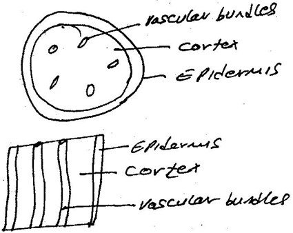 (i) Make a labeled plan diagram of the section
(ii) From your observations of the section, to which class does the specimen belong?
Class Dicotyledonous – rej. Dicot and cotyledon
Reason Vascular bundles arranged in a ring/ circle/ vascular bundles is on either side of pith/distinct cortex.